
Immunohistochemistry (IHC) is used to detect and localize antigens (e.g., proteins) in cells or tissue sections by exploiting the principle of antibodies binding specifically to antigens in biological tissues.
ChemPartner has accumulated extensive hands-on experience in histopathology that provides support to research in oncology, immunology, inflammation, neuroscience, metabolic and cardiovascular diseases, toxicology, and the discovery of large and small molecule drugs.
ChemPartner is dedicated to providing high-quality and efficient services for IHC assay development, protocol validation, single/double staining, image collection, and data analysis/interpretation. For non-commercial antibodies, ChemPartner also provides full assay development for validation and optimization before formal study.
- Equipment: IHC autostainer (Dako AS Link48), digital image scanner (Aperio ScanScope XT ss001310), Nikon Image Collection Microscope, etc.
- Data analysis software: ImageScope (Aperio), Image Pro Plus, Image J, GraphPad Prism 5, etc.
- Antibodies with established methods: already established IHC methods for 110+ antibodies
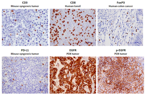
- Oncology: Ki67, EGFR, p-EGFR, HER2, HER3, HER4, MEK1/2, Akt, p-Akt, ERK, p-ERK, PTEN, HGF, IGFR, c-Met, p-c-Met, c-Myc, n-Myc, E-Cadherin, p-Cadherin, ER, CD31, HIF 1 alpha, Cleaved Caspase-3, VEGF, etc.
- Immunology: CD3, CD4, CD8, Foxp3, PD-1, PD-L1, CTLA-4, Iba-1, MPO, etc.
- Neuroscience: Tyrosine hydroxylase, MBP, MOG, Doublecortin, NG2, GFAP, etc
- Tissue types: FFPE and frozen samples of cells and tissue, tissue microarray (TMA)
Representative Images of IHC staining
Representative Images of IHC Double Staining
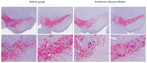
- TH(Pink): Tyrosine Hydroxylase, Millipore, AB152, Rabbit IgG; α-Synuclein Phospho (Ser129)(Brown): Biolegend, #825701, Mouse IgG2a
- Blue arrows indicate the positive cells of co-staining; Phosphorylation of α-Synuclein Ser-129 is selective and extensive in synucleinopathy lesions
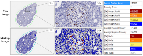
Image Analysis with Aperio Quantification software
Deliverable:
- Raw data together with analyzed data in MS Excel format
- Ordinary images in JPEG format or scanned images in SVS format
- Project update and summary in MS PowerPoint format
- Final report in MS Word format (including purpose, methods, results, and data analysis)
Sample Submission:
- Test samples could be submitted
- FFPE blocks of tissues or cells
- Oct-embedded samples
- Tissues and cells fixed in formalin
- Fresh frozen samples