免疫组织化学 (IHC)利用抗体与生物组织中的抗原特异性结合的原理,用于检测和定位细胞或组织切片中的抗原(例如,蛋白质)。
ChemPartner 在免疫组织化学方面积累了丰富的实践经验,为肿瘤、免疫、炎症、神经、代谢和心血管疾病的研究提供支持,同时也支持毒理研究以及大分子和小分子药物的发现。
ChemPartner 致力于为 IHC 检测开发、方案验证、单/双染色、图像采集和数据分析/解读提供优质高效的服务。 对于非商业抗体,ChemPartner 还提供完整的方法开发服务,在正式研究之前完成方法验证和优化。
- Equipment: IHC autostainer (Dako AS Link48), digital image scanner (Aperio ScanScope XT ss001310), Nikon Image Collection Microscope, etc.
- Data analysis software: ImageScope (Aperio), Image Pro Plus, Image J, GraphPad Prism 5, etc.
- Antibodies with established methods: already established IHC methods for 110+ antibodies
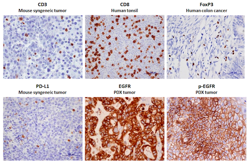
- Oncology: Ki67, EGFR, p-EGFR, HER2, HER3, HER4, MEK1/2, Akt, p-Akt, ERK, p-ERK, PTEN, HGF, IGFR, c-Met, p-c-Met, c-Myc, n-Myc, E-Cadherin, p-Cadherin, ER, CD31, HIF 1 alpha, Cleaved Caspase-3, VEGF, etc.
- Immunology: CD3, CD4, CD8, Foxp3, PD-1, PD-L1, CTLA-4, Iba-1, MPO, etc.
- Neuroscience: Tyrosine hydroxylase, MBP, MOG, Doublecortin, NG2, GFAP, etc
- Tissue types: FFPE and frozen samples of cells and tissue, tissue microarray (TMA)
Representative Images of IHC staining
Representative Images of IHC Double Staining
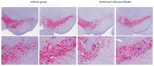
- TH(Pink): Tyrosine Hydroxylase, Millipore, AB152, Rabbit IgG; α-Synuclein Phospho (Ser129)(Brown): Biolegend, #825701, Mouse IgG2a
- Blue arrows indicate the positive cells of co-staining; Phosphorylation of α-Synuclein Ser-129 is selective and extensive in synucleinopathy lesions
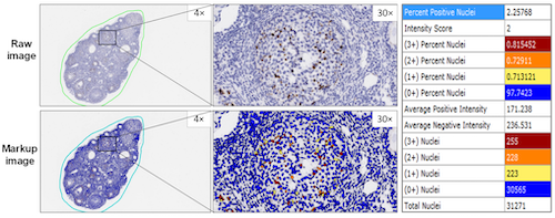
Image Analysis with Aperio Quantification software
交付成果:
- 原始数据和 MS Excel 格式的分析数据
- JPEG 格式的普通图像或 SVS 格式的扫描图像
- MS PowerPoint 格式的项目更新和总结
- MS Word 格式的最终报告(包括目的、方法、结果和数据分析)
样品提交:
- 可以提交测试样品
- FFPE 组织或细胞块
- Oct包埋的样本
- 在福尔马林中固定的组织和细胞
- 新鲜冷冻样品
