

Mixed Lymphocyte Reaction (MLR) Assay
The MLR assay is considered the ‘gold standard’ T cell functional assay for the test of T cell immune checkpoint inhibitors Dendritic cells co-cultured with allogeneic CD3+ T cells Readouts: IFN-γ and IL-2 production Suitable to test both antibodies and small...


Whole Blood/PBMC Stimulation and Cytokine Release Assay
LPS-induced Cytokine Release in Human PBMC


Primary Immune Cell In Vitro Assays
Whole Blood/PBMC Stimulation and Cytokine Release Assay Mixed Lymphocyte Reaction (MLR) Assay CMV Antigen Recall Assay Tumor/T Cell Co-culture Assay T Cells (Total, CD4+, CD8+) Activation and Proliferation Assays Th Subsets Differentiation (Th1/2/17/Treg) Assays Treg...


Case Studies in the Syngeneic Model
Efficacy Test in CT2.WT Model Tumor Growth Survival Evaluation (Endpoint: TV > 2000mm3 Mice under combination therapy exhibited survival benefit Efficacy Test in 4T1 Resection Model Tumors were resected on D13; Half mice to observe survival;...


Case Studies in PDXs
Mouse Clinical Trial in PDXs Objective: to evaluate drug efficacy across 17 colorectal PDXs Pre-defined Clinical Endpoints Tumor Progression A tumor volume change >73% by the end of treatement Tumor Stasis A tumor volume change between -66% and 73% by the end of...


Model Generation Study in CDXs
Tumor Volume Change of NCl-H661 subQ Model Total Flux in U87MG-Luc2 Orthotopic Model


Case Studies in CDXs
Efficacy Study in CDXs Tumor Growth Inhibition in MV-4-11 Survival Rate in A375 Brain Inoculation Tumor Growth Inhibition in Hep3B 2.1-7 Efficacy and Biomarker Assessment in CDXs Cancer Discovery. 2018 Jul;8(7):836-849 Cell line derived allograft and xenograft models...


Molecular Pathology: Immunofluorescence (IF)
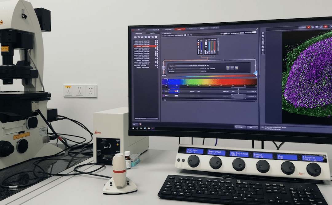
Immunofluorescence (IF) staining is used to detect and localize antigens (e.g., proteins) in cells or tissue sections by using of fluorescent dye-conjugated antibodies or non-antibody methods of fluorescent staining (eg. DAPI). Compared with immunohistochemistry...


